

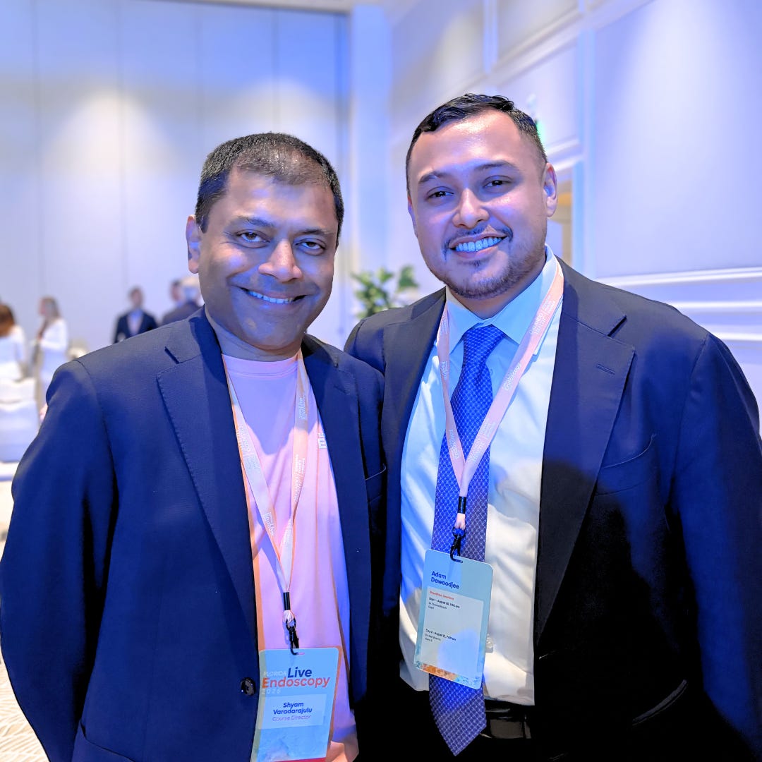
From High-Risk Novelty to Illuminated Precision
In 1868, looking inside a living human stomach was an act of raw terror. German physician Adolf Kussmaul peered down a patient’s throat using a rigid metallic tube inspired by the reckless anatomical feats of traveling sword swallowers. It was brutal, high-risk, and deeply traumatic, prompting the global medical community to hit pause on internal visualization for decades. Contrast that historical nightmare with the seamless reality of a modern high-definition endoscopy suite, where crystal-clear monitors render mucosal tissue in breathtaking topographical detail.
Yet, medical milestones are rarely forged by sterile corporate breakthroughs alone. As explored through the investigative lens of Surgery Gets Smarter, true progress is driven by relentless human ingenuity, makeshift engineering, and cross-generational grit across a 150-year journey.
By connecting the post-WWII garage-lab grit of early gastrocamera inventors to legends like Dr. Varadarajulu and the global faculty at Florida Live Endoscopy 2026, we see an unbroken lineage of innovation. It is a shared mission aimed at a single, unchanging goal: turning blind, aggressive surgery into illuminated, curative precision without punishing the human body.
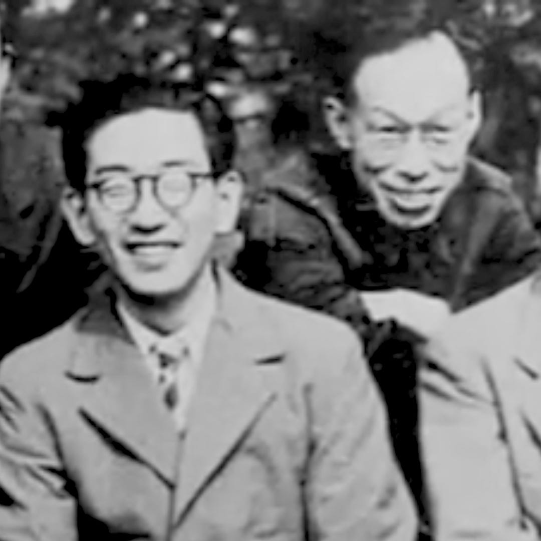
Dr. Uji and the Gastrocamera Breakthrough
This spirit of relentless tinkering took root in post-war Tokyo, where Dr. Tatsuro Uji teamed up with Olympus engineers Mutsuo Sugiura and Shoji Fukami during a time of severe resource scarcity. Self-funding their research out of their own pockets, they set up a makeshift garage lab to build the world’s first gastrocamera. They replaced rigid, dangerous metal tubes with a soft, flexible 12 mm vinyl pipe. To capture internal photos, they improvised apparatuses to hand-slice standard 35 mm film down to tiny 6 mm strips, paired with a custom-crafted 5 mm double-filament lamp wired to a tiny camera mechanism inside the tip. Delivering a working prototype in just four months, they captured the world’s first clear photographs inside a living stomach. From Tokyo basements to European operating theaters, these pioneers established that therapeutic safety could triumph over procedural trauma.
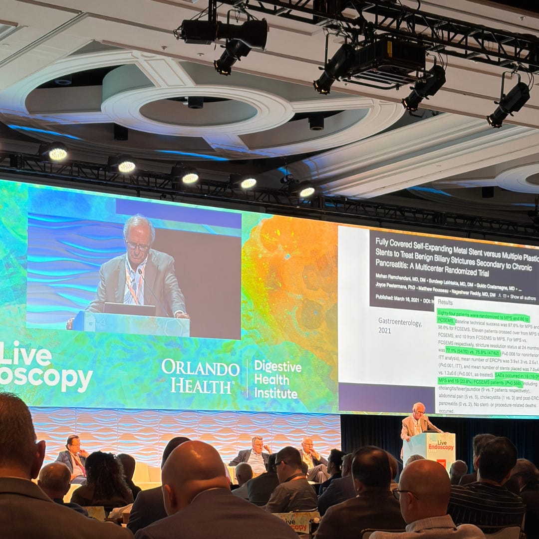
How Fiber-Optics Replaced the Scalpel
To understand how far gastroenterology has come, one must look back at the baseline of the mid-20th century. Before flexible optics, internal intervention meant large surgical incisions and significant patient risk. Clinicians had to rely heavily on indirect diagnostic clues, while early endoscopic tools were widely dismissed by traditionalists as impractical novelties relegated to storage rooms.
Overcoming widespread skepticism required both fundamental optical physics and clinical courage. Innovators like Harold Hopkins revolutionized physics with coherent fiber bundles, while Basil Hirschowitz transformed those principles into the first operational flexible fiberscopes. These technical breakthroughs allowed a daring generation of early endoscopists to bridge the gap between academic theory and daily procedural care.
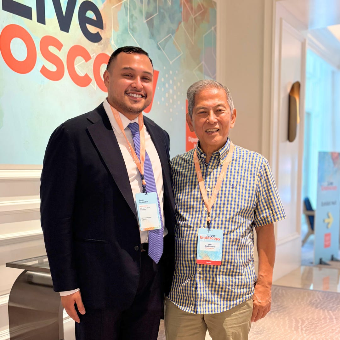
Dr. Nib Soehendra’s Endoscopic Revolution
Between 1968 and 1972, a seismic diagnostic revolution swept through medicine, introducing esophagoscopy, colonoscopy, and ERCP. Yet, looking inside was only the first step. The true revolution occurred when visionaries transformed endoscopes from simple viewing instruments into active surgical workshops.
Chief among these therapeutic pioneers was Dr. Nib Soehendra, whose work in Hamburg fundamentally reshaped the field. Confronted with life-threatening gastrointestinal hemorrhages and impassable biliary obstructions, Soehendra refused to accept open surgery as the only recourse. He pioneered endoscopic sclerotherapy to halt acute variceal bleeding and invented the mechanical lithotripter, allowing physicians to crush massive gallstones inside the duct without making a single external incision.
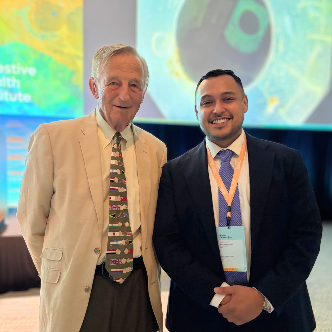
Meeting Dr. Peter Cotton at Florida Live 2026
Moving from early historic prototypes to the high-stakes arena of Florida Live Endoscopy 2026 brings this lineage into sharp focus. Here, the foundational work of icons like Dr. Peter Cotton, who helped introduce ERCP to Western medicine and executed landmark sphincterotomies in the 1970s, culminates in live-case masterclasses.
Watching modern experts navigate intricate biliary tracts and resect early-stage lesions on high-definition displays highlights how far global collaboration has advanced the discipline. Yet the core principle remains unchanged: detect and eradicate disease early while protecting the patient.
Furthermore, the evolution of the modern suite is as much about human collaboration as optical engineering. Just as early endoscopy required close partnership between clinicians and specialized nurses, today’s live demonstrations succeed because cohesive, multidisciplinary teams work in unison.
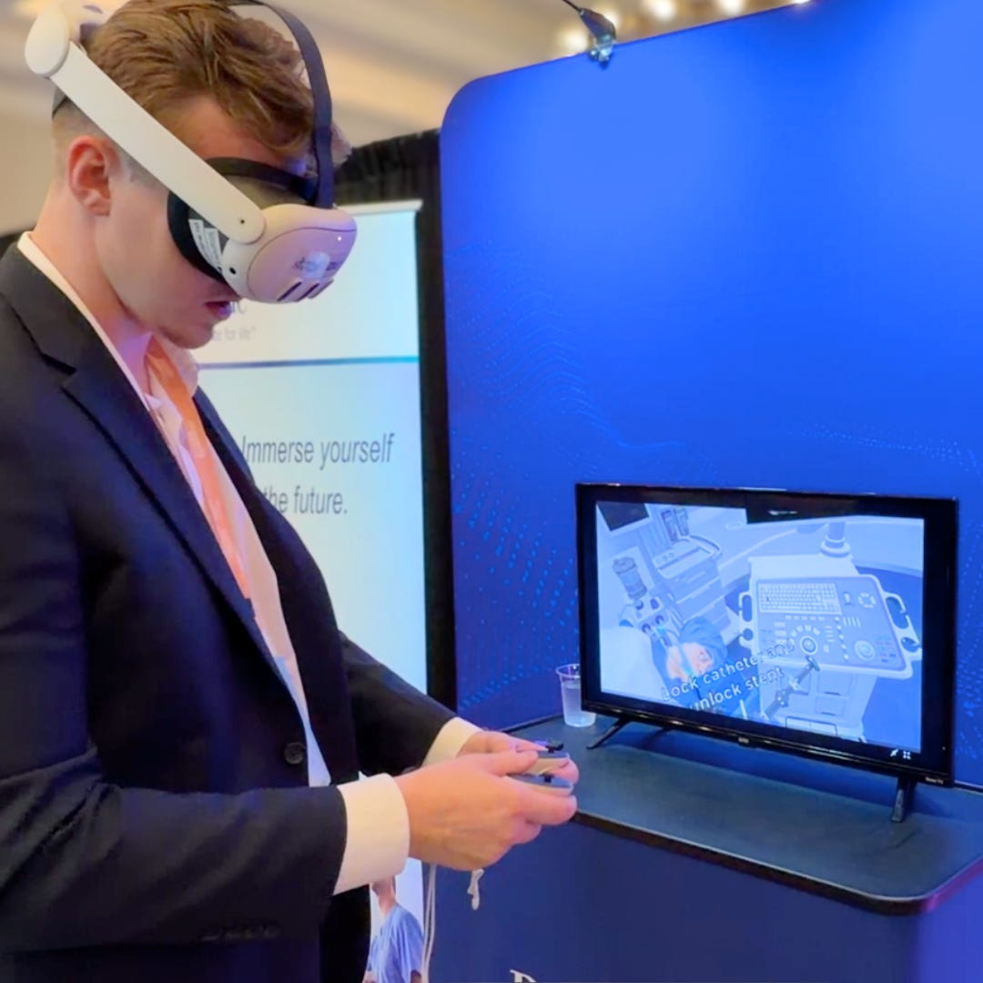
V.R. and the High Tech O.R. Suites of Tomorrow
As we look toward the horizon of modern MedTech, marked by virtual reality training simulators, artificial intelligence integration, and advanced narrow-band imaging, we encounter a familiar industry challenge. It is tempting to assume that progress requires increasingly costly, complex machinery that risks pricing many healthcare systems out of advanced care.
True surgical innovation, however, follows a broader philosophy. Whether watching a pioneer refine a stone-crushing snare or observing a clinician hone their dexterity on a cutting-edge VR simulator, the primary objective remains constant: identify clinical pain points, reduce ergonomic strain on the operator, and expand access to gentle, precise treatment. Technology achieves genuine intelligence only when our commitment to reducing trauma matches the clarity of the image on screen.
Learn More & Resources
To dive deeper into the historical evolution of gastrointestinal endoscopy, early flexible optics, and the origins of modern therapeutic ERCP, explore these resources online:
Read Dr. Peter Cotton’s retrospective on fifty years of endoscopic retrograde cholangiopancreatography and the evolution of pancreaticobiliary care through Gastrointestinal Endoscopy.
Explore the seminal 1957 paper by Basil Hirschowitz and colleagues detailing the design and testing of the first operational flexible fiberoptic endoscope through the University of Michigan Medical Bulletin.
Review the historical arc of digestive endoscopy from 19th-century rigid visualization to late-20th-century electronic optics through ResearchGate.
Watch the historical discussion featuring Japanese Ambassador Akio Hosono and gastroenterologist Dr. Julio Murra-Saca through YouTube.
References
Cotton, P. B. Fifty years of ERCP: a personal review. Gastrointestinal Endoscopy. published August 2018.
Hirschowitz, B. I., Peters, C. W., & Curtiss, L. E. Preliminary report on a long fiberscope for examination of stomach and duodenum. University of Michigan Medical Bulletin. published May 1957.
Niwa, H. The History of Digestive Endoscopy. Digestive Endoscopy. published January 1996.
Soehendra, N., & Reynders-Frederix, V. Palliative bile duct drainage: An endoscopic method. Endoscopy. published January 1980.
Murra-Saca, J. (2008, January 30). The Untold History of The Gastrointestinal Endoscopy Part 2 (2/6) [Video].YouTube; Dr.Julio Murra Saca Endoscopia El Salvador.